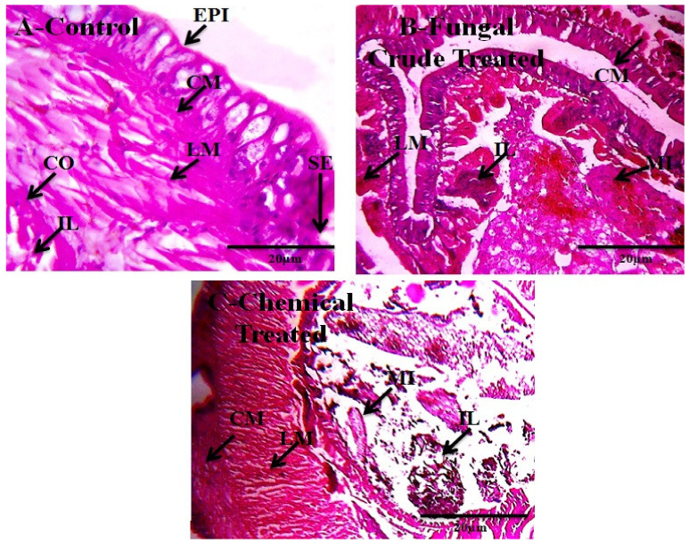
Figure 3.
The M. anisopliae secondary metabolites (200 µg/mL) were exposed E. eugeniae and after 30 days of treatment, the earthworm gut tissues were sectioned for histopathological evaluation and magnified at 40× under a light microscope. (A) is control (without fungal crude extract treatment); (B) is fungal secondary metabolites treated; and (C) is Monocrotophos 200 ppm/kg treated. In the control and entomopathogenic fungi crude extract treatments, no changes were observed, but chemical pesticide treatment of several gut tissues morphology and shapes changed in the lumen tissues was entirely spoiled compared with control (EPI-epidermis, SE-setae, IL-intestinal lumen, LM-longitudinal muscle, CO-coelom, CM-circular muscle, MI-mitochondrion).

