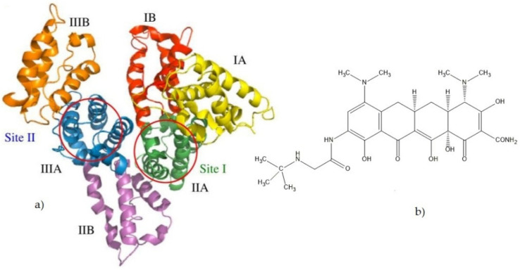
Figure 1.
(a) Crystal structure of HSA. The subdivision of HSA into domains (I-III) and subdomains (a,b) is indicated, and the approximate locations of site I and site II are shown. Atomic coordinates were taken from the PDB entry 1UOR. The illustration was performed with PyMOL [4] (b). Chemical structure of tigecycline.

