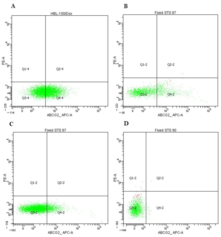
Figure 5.
ABCG2 expression analysis by cytofluorometry. (A) HBL-100/Dox cells were stained with APC-labelled anti-ABCG2 antibodies (ABCG2 expressed by 50% cells). Dox-resistant cell subline HBL-100/Dox with overexpressed ABCG2 (BCRP) was obtained from the HBL-100 cells after the selection with Dox. HBL-100/Dox cells were used as positive control for the determination of ABCG2 staining. (B) Expression of ABCG2 in 17.1% of cells in STS 87 (liposarcoma). (C) Expression of ABCG2 in 8.6% of cells in STS 97 (undifferentiated pleomorphic sarcoma). (D) Expression of ABCG2 in 0.4% of cells in STS 90 (synovial sarcoma).

