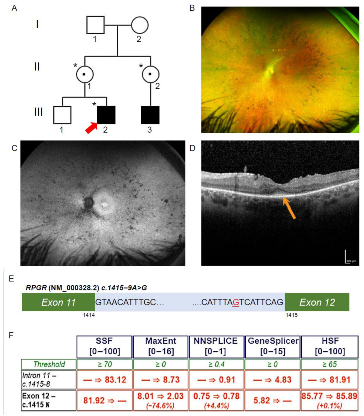
Figure 1.
Ophthalmic investigations and RPGR novel variant. (A) The proband (Patient III-2; red arrow) was affected with RCD, as was his maternal cousin (III-3). The proband was hemizygous for the intronic variant in RPGR (*), and his mother and maternal aunt were heterozygous for the same variant (*), while his unaffected brother did not have the variant. Known female carriers are denoted (•). (B) Patient III-2 fundal images demonstrating mild pigmentary disturbance in the mid periphery. (C) Ultra-widefield fundus autofluorescence demonstrating a narrow ring of hyperautofluorescence around the fovea and patchy hypoautofluorescence scattered throughout the fundus. (D) The Spectral Domain Optical Coherence Tomography demonstrated a residual blurred ellipsoid zone at the fovea (orange arrow). (E) Patient III-2, RPGR, c.1415 − 9A>G highlighted in red in intron 11 of RPGR (NM_000328.2). (F) In silico prediction across five programs using Alamut Visual scored above the threshold for the novel variant c.1415 − 9G>A behaving as an acceptor site. Scores were below threshold for the canonical acceptor site, indicating a potential loss of function in the presence of the novel variant.

