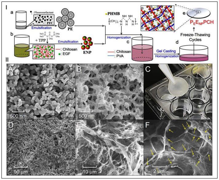
Figure 1.
Analyses of the structure and morphology of the PEENPPCH. (I) Schematic diagram of the PEENPPCH fabrication procedures, including the manufacture of PEs and ENPs (a,b), hetero-composite hydrogel formulation (c), casting (d), and formation after eight freeze–thaw cycles (e). (II) Photomicrographic images of the manufactured nanoparticles and hetero-composite hydrogel. (A,B) SEM images of the ENPs (A) and PEs (B) at 50,000× magnification. (C) Photograph of the real PEENPPCH sample. (D–F) SEM images showing the surface of the PEENPPCH at 500× magnification (D), the inner structure of the PEENPPCH at 2000× magnification (E), and the inner structure of the PEENPPCH at 10,000× magnification (F). Arrows indicate the nanoparticles (ENPs and PEs) adhered to the polymeric fibers.

