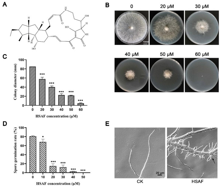
Figure 1.
HSAF caused growth defect in N. crassa. (A) Chemical structure of HSAF; (B) Growth of N. crassa on Vogel’s plates supplemented with HSAF (0, 20, 30, 40, 50, 60 µM) for 36 h. Images show the results of one out of three experiments, and the statistical analysis of colony diameter is shown in (C); (D) Germination rate of N. crassa under HSAF treatment. Asterisks indicate significant differences (*, p < 0.05; ***, p < 0.001); (E) Microscopic morphology of N. crassa grown on Vogel’s media without (CK) or with 30 µM HSAF treatment (HSAF) for 6 h. The black arrow indicates the hyper-branch.

