
Fig. 1.
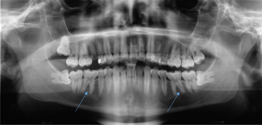
Routine X-Ray taken in January 2019 by the primary dentist. Tooth 36 and 46 without any apical osteolysis before the intensive of bruxism started. Both teeth have no signs of caries and no fillings are visible
Official websites use .gov
A
.gov website belongs to an official
government organization in the United States.
Secure .gov websites use HTTPS
A lock (
) or https:// means you've safely
connected to the .gov website. Share sensitive
information only on official, secure websites.
Routine X-Ray taken in January 2019 by the primary dentist. Tooth 36 and 46 without any apical osteolysis before the intensive of bruxism started. Both teeth have no signs of caries and no fillings are visible