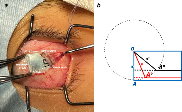
Fig. 1.
The procedure of slanted recession of LR. a Taken during the surgery, recession 4 mm of the upper pole and recession 6 mm of the lower pole. The slanted amount is 2 mm. b The diagram of slanted recession procedure. O is the opper pole of LR. OA (blue) is the new insertion and a (blue) is the central point of OA in the standard recession procedure. During the procedure in our study, the scleral pass of the upper pole of LR was firstly performed and then the scleral pass of the lower pole. OA’ (red) is the slanted insertion and a’ (red) is the central point of OA’ in the 1 mm slanted recession procedure. OA” (black) is the slanted insertion and a” (black) is the central point of OA” in the 2 mm slanted recession procedure. To keep the original length of LR insertion, the actual width of LR, perpendicular to the direction of the muscle force, is narrowed and the central point of new insertion is shifted upwards a bit

