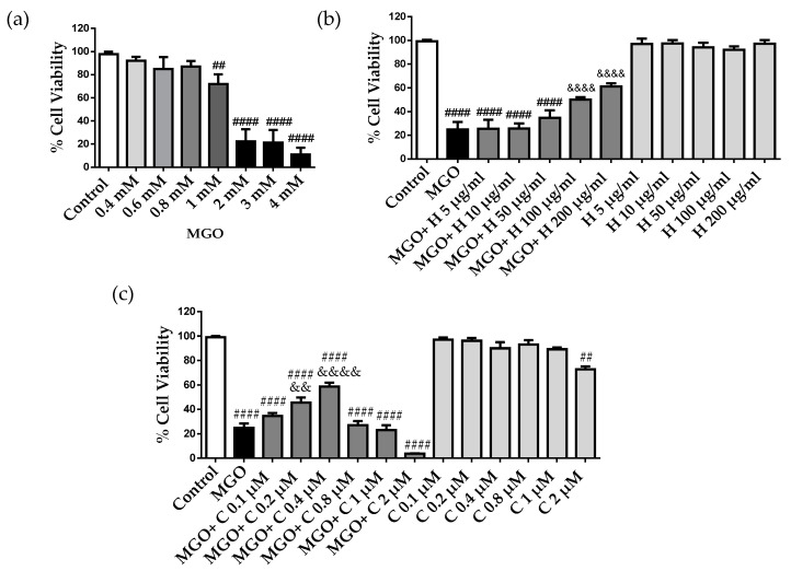
Figure 2.
MGO cytotoxicity in bEnd3 cerebral endothelial cells, effect of rHDLs (H) and curcumin (C). (a) The cells were treated with different MGO concentrations (0.4–4 mM) for 24 h. (b) The cells were pre-treated with different concentrations of rHDLs, 5–200 μg/mL, (H) and (c) curcumin (C), 0.1–2 μM, for 1 h, followed by the addition of 2 mM MGO for 24 h. Cell viability was assessed by the MTT assay. Data are presented as mean ± SD of three independent experiments (n = 3). ## p ˂ 0.01, #### p ˂ 0.0001 as compared to control. && p ˂ 0.01, &&&& p ˂ 0.0001 as compared to MGO.

