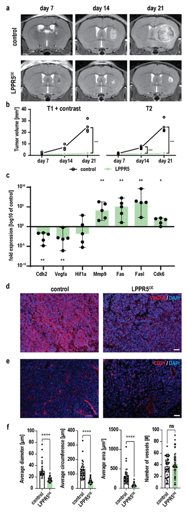
Figure 3.
In-vivo characterization of LPPR5OE tumors. (a) Coronal T2 MR images of tumor baring mice 7-, 14- and 21-days post tumor cell injection of control GL261 and LPPR5OE clones. (b) T1 contrast and T2 MR image quantification shows higher sensitivity for volumetry with T2 sequences in these tumors [T1+contrast; day 21 *** p = 0.0080, T2; day 14 *** p = 0.0045, day 21 *** p = 0.0097, mixed effect analysis, Sidak’s multiple comparisons test]. (c) qPCR expression screening of apoptotic and angiogenic genes in LPPR5OE tumors compared to control tumors [median and range of the values is plotted as whiskers. ** p = 0.008, * p = 0.032, Mann-Whitney U-Test with independent sampling]. (d) Immunohistochemical staining of Vegfa in control and LPPR5OE tumors using equal exposure settings [scale bar represents 50 µm]. (e) Immunohistochemical staining of blood vessels (CD31) in control and LPPR5OE tumors [scale bar represents 50 µm]. (f) Automated Analysis of blood vessel parameters show significant different diameter (**** p < 0.0001), vessel circumfluence, edge length (**** p < 0.0001), and area (**** p < 0.0001). No difference was found in the total number of vessels. ns = non significant.

