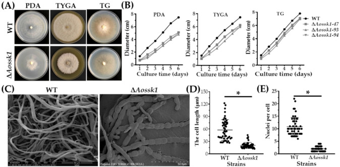
Figure 1.
Comparison of mycelial growth, morphology, and cell nuclei between WT and ΔAossk1 mutant strains. (A) Colony morphologies of WT and ΔAossk1 mutant strains cultured on PDA, TG, and TYGA plates for 6 days at 28 °C. (B) Colony diameters of WT and ΔAossk1 mutant strains cultured on PDA, TYGA, and TG plates. (C) Mycelial morphologies of WT and ΔAossk1 mutant strains were observed by scanning electron microscopy. (D) Comparison of mycelial lengths of WT and ΔAossk1 mutant strains. (E) Comparison of cell nuclei in the hyphae of WT and ΔAossk1 mutant strains. An asterisk (D,E) indicates a significant difference between ΔAossk1 mutant and WT strain (n = 50, Tukey’s HSD, p < 0.05).

