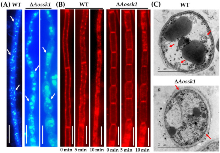
Figure 3.
Comparison of autophagy and endocytosis between WT and ΔAossk1 mutant strains. (A) Comparison of autophagosomes in the hyphal cells of the WT and ΔAossk1 mutant strains. White arrows: autophagosomes. Bar = 10 μm. (B) Endocytosis in the WT and ΔAossk1 mutant strains at different times; Bar = 10 μm. (C) Observation of autophagic vacuole in hyphal cells using transmission electron microscopy. Red arrows: autophagic vacuole.

