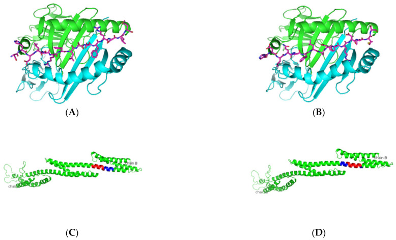
Figure 5.
Predicted α-fodrin peptide binders docked on HLA-DRB1*0103. (A,B) Based on the prediction by SMM-align using IEDB, the top two peptides with IC50 values of 12 were docked onto the crystal structure of HLA-DRB1*0103 PDB structure 1A6A, and the most optimum predicted position of docking is indicated for both peptides SHDLQRFLSDFRDLM and HDLQRFLSDFRDLMS with the core sequence of SHDLQRFLS and FLSDFRDLM, respectively. (C,D) The highlighted region indicates the presence of the peptides in the three-dimensional structure of the α-fodrin protein.

