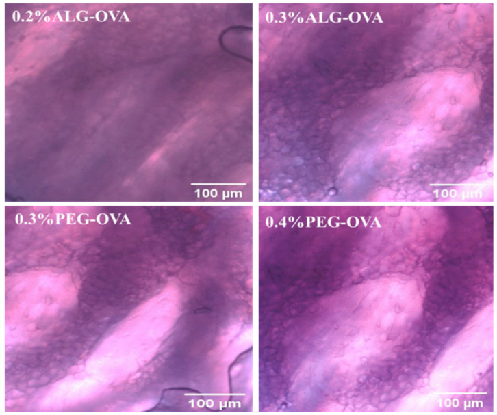
Figure 8.
Fluorescence microscopy images showing the mucoadhesion of unlabeled ALG- and PEG-coated chitosan nanoparticles (control) to the pig oral mucosa. Extracted images were analyzed using the Image J software in red to green (R/G) intensity and no green fluorescence was observed as a distinguishing feature for the samples compared with Figure 9 below.

