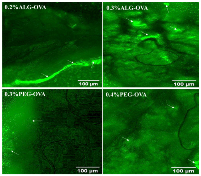
Figure 9.
Fluorescence microscopy image showing the mucoadhesion of FITC-labelled ALG- and PEG-coated chitosan nanoparticles to the pig oral mucosa (white arrows show areas of intense green fluorescence on the mucosa where nanoparticles were resident). The fluorescence images were analyzed to extract the green intensities using the Image J software. This was used to determine nanoparticle presence (arrow bar) on the pig mucosa tissue. The images obtained for 0.2% ALG-OVA nanoparticles showed a progressive increase in the intensity of green fluorescence emitted from the FITC-labelled particles of this formulation on pig mucosa tissue compared to the 0.3% ALG-OVA. This indicates that the 0.2% ALG-coated particles could potentially be more adhesive and possess a higher binding efficacy than the 0.3% ALG-coated particles. In the case of the PEG-coated samples, the 0.3% PEG-modified chitosan nanoparticles showed higher green fluorescence intensities and therefore better adhesion than the 0.4% PEG-modified nanoparticles.

