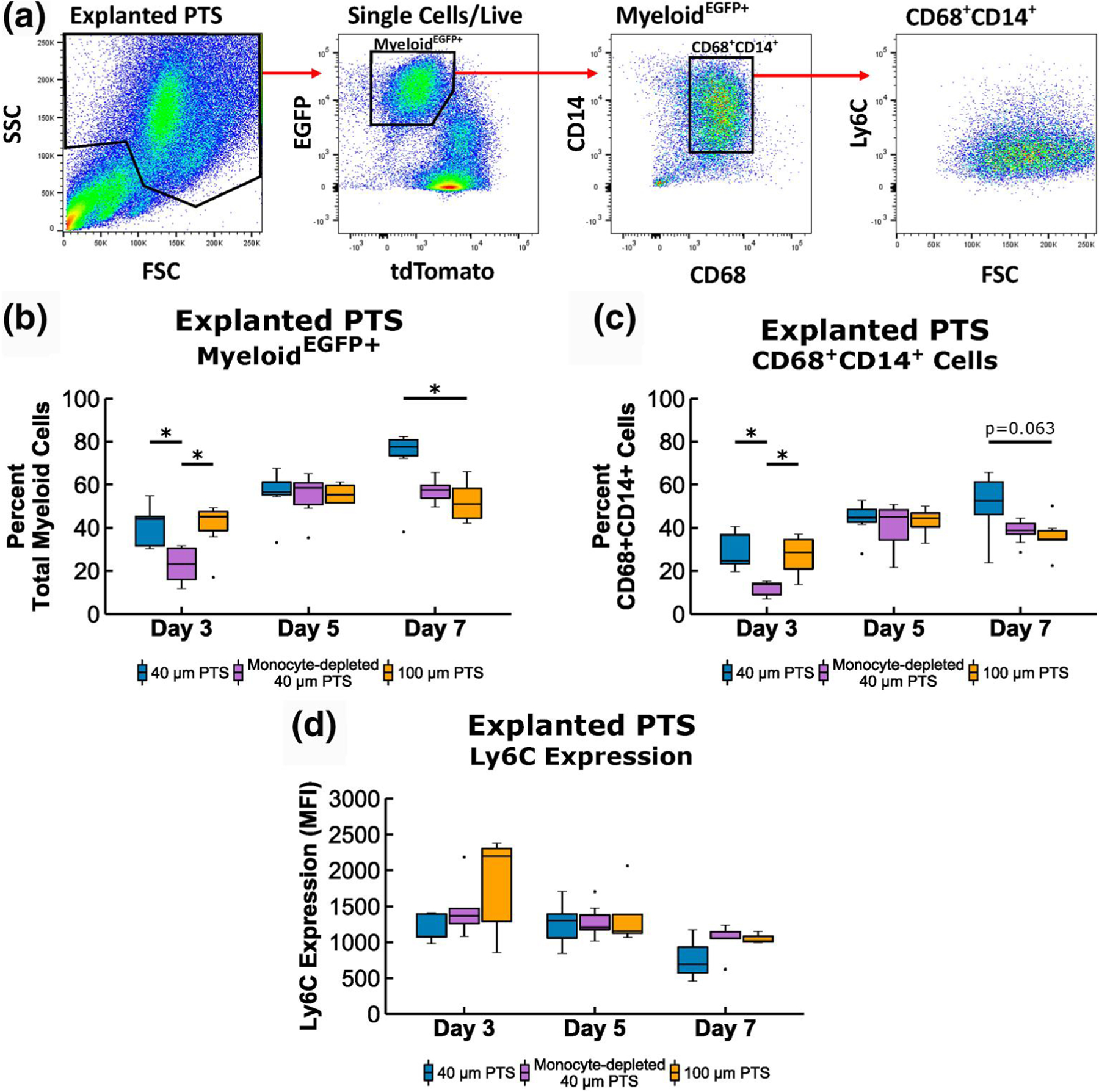
FIGURE 2.

Flow cytometric analysis of scaffold resident cells recovered from 40 μm precision-templated scaffolds (PTS) or 100 μm PTS implanted subcutaneously in LysM-Cre+/0:mT/mG+/0 mice, and from 40 μm PTS implanted subcutaneously in monocyte-depleted LysM-Cre+/0:mT/mG+/0 mice. (a) Explanted scaffold-resident cells were gated on MyeloideGFP+ expression, and the monocyte linage was further discriminated by CD68+CD14+ expression. Cell aggregates, doublets, and dead cells were excluded from analysis. (b) Percentage of total myeloid cells from explanted PTS. (c) Percentage of MyeloideGFP+ CD68+CD14+ cells from explanted PTS. (d) median fluorescent intensity (MFI) of Ly6C expression from explanted PTS. * denotes p < 0.05 and ** denotes p < 0.01; n = 4–8 animals per group
