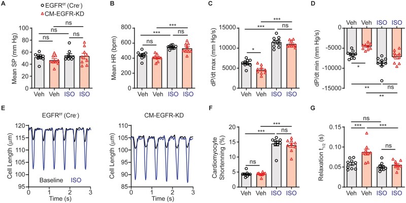
Figure 5.
Cardiomyocyte-specific EGFR downregulation impairs relaxation dynamics. Haemodynamic evaluation of mean systolic pressure (SP, A), mean heart rate (HR, B), +dP/dt max (C) and -dP/dt max (D) in adult EGFRf/f (Cre−) vs. CM-EGFR-KD mice at baseline and in response to infusion with 10 ng ISO, n = 9 each. *P < 0.05, **P < 0.01, ***P < 0.001, ordinary one-way ANOVA with Tukey’s multiple comparisons test. (E) LV cardiomyocytes were freshly isolated from adult EGFRf/f (Cre−) vs. CM-EGFR-KD hearts and cardiomyocyte length measured at baseline (black line) and after stimulation with ISO (0.1 μM, red line), as shown in tracings representative of n = 9 individual cardiomyocytes isolated from n = 3 mice per genotype, with quantification of cardiomyocyte shortening (F) and time to 50% relaxation (G) summarized in histograms. ns, not significant, ***P < 0.001, ordinary one-way ANOVA with Tukey’s multiple comparisons test.

