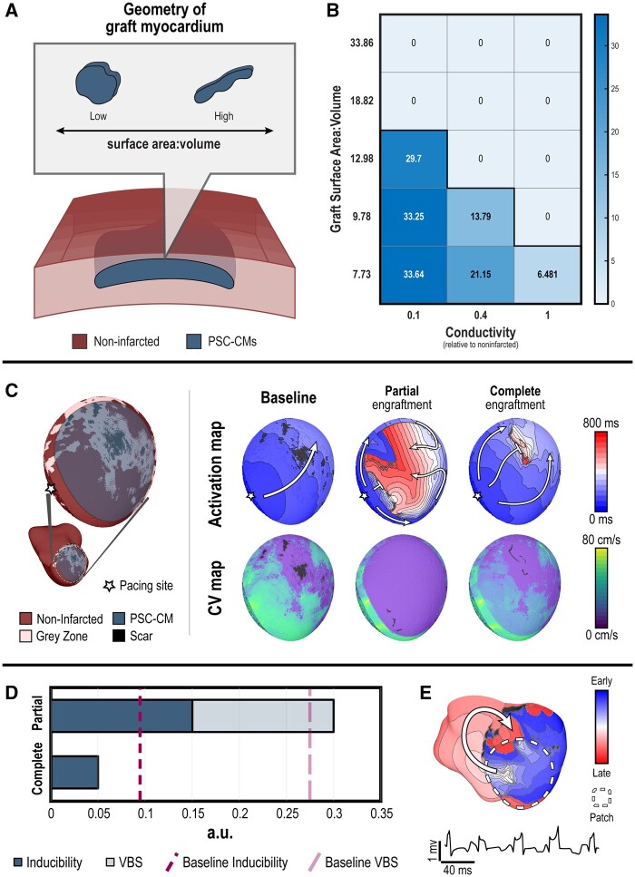
Figure 3.
A re-entrant VT mechanism dominates over a focal mechanism in patch remuscularization, but partial engraftment of PSC-CM patches exacerbates VT burden due to conduction slowing through the patch. (A) Schematic of experimental set-up. The emergence and rate of focal ectopic propagations from graft myocardium was observed for PSC-CMs embedded within a cut-out of the ventricular myocardial wall; the surface area-to-volume ratio of the graft was altered. (B) Frequency (b.p.m.) of graft-induced focal ectopic propagations across different graft geometries (i.e. surface area: volume ratio) and conductivities. (C) Remuscularization with a PSC-CM patch (radius = 3.2 cm) applied to segment of infarct (L1) along the anterior, inferior LV of P1 (left inset) was simulated; simulated pacing occurred at the starred site. Electrical propagation across the patch was observed under baseline (left), partial engraftment (middle), and complete engraftment (right); activation maps (top) and conduction velocity maps (bottom) indicate the sequence of epicardial electrical activation and local conduction velocity, respectively. (D) Bar graph of inducibility (dark blue) and VT burden (VBS, light blue) for P1 under conditions of partial and complete engraftment; dashed lines indicate inducibility (dark pink) and VT burden (light pink) at baseline. (E) Activation map and pseudo-ECG of the single emergent VT morphology induced in P1 when complete engraftment was simulated.

