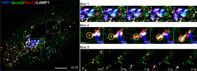
Fig. 5.
Secretory and degradative procollagen export from ERES. Left panel, first frame of 1 s/frame time-lapse image series (Movie 3) of a Col1a2GFP-F cell transfected with mTagBFP2-ARF1, mCherry-Sec23, and LAMP1-Halo. Right panels, selected time-lapse frames of areas marked by boxes 1–3 in the left panel. Box 1 frames show a transport vesicle delivering procollagen from ERES to Golgi (encircled in cyan) and a lysosome (encircled in magenta). Box 2 shows ERES being engulfed by a lysosome (encircled in yellow). Box 3 shows lysosome (encircled in magenta) movement and fusion with a quasi-stationary vesicular structure (likely a lysosome as well). Fast mode (1 s/frame), 4-color, super-resolution Airyscan imaging was necessary for visualizing and distinguishing rapidly moving procollagen ER–Golgi intermediates and lysosomes. Some crosstalk between fluorescence channels in this imaging mode was unavoidable, causing ambiguity in interpretation of weaker fluorescence signals (e.g., the vesicular structure in Box 3). Scale bars = 10 µm (whole cell) and = 1 µm (zoomed regions)

