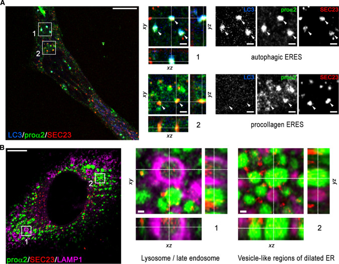
Fig. 6.
Procollagen sorting at ERES. a Super-resolution (Airyscan) images of procollagen ERESs marked by GFP-proα2(I) and COPII coat protein Sec23 (box 2, arrowheads) and autophagic ERES structures colocalized with autophagy marker LC3 (box 1, arrowheads) in a Col1a2GFP-A cell transfected with mCherry-Sec23 and mTagBFP2-LC3 in the presence of Asc2P. b Airyscan images of an autophagic procollagen ERES surrounded by lysosomal membrane marked with LAMP1 (box 1) and multiple vesicle-like procollagen pools in dilated ER studded with Sec23 (region 2) in a Col1a2GFP-A cell transfected with mTagBFP2-Sec23 and LAMP1-Halo. The cells were treated for 8 h with leupeptin and 3 h with brefeldin A before being fixed, to prevent procollagen export from the ER and rapid degradation of Sec23 in lysosomes [19]. It is noted that the Sec23 signal inside the lysosome is just as strong as at ERESs (outside the lysosome), while the procollagen signal is fainter due to reduced GFP fluorescence at acidic pH [63]. Scale bars in a and b = 10 µm (whole cell) and = 1 µm (zoomed regions)

