Fig. 7.
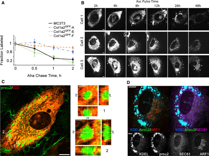
Procollagen trafficking and accumulation in the ER. a Kinetics of procollagen clearance from cells measured by pulse chase labeling with Aha that replaces Met in proteins synthesized during Aha pulse in Met-free media. Plotted is the fraction of Aha-labeled chains relative to the start of Aha chase in Aha-free, Met-containing media. The measurements of procollagen clearance kinetics were performed in the same experiments as measurements of secretion kinetics described in Fig. 4c. Error bars show SEM based on the same number of replicates as in Fig. 4c. b and Movie 4 Time-lapse images of procollagen clearance, formation of procollagen pools, and dynamics of these pools in the ER of Col1a2GFP-A cells after addition of Asc2P. Cells 1–3 illustrate different observed behaviors, including formation and slow clearance of large procollagen pools in the ER (cell 1), no formation of such pools (cell 2), and accumulation and enlargement of dilated ER containing procollagen (cell 3). c Airyscan imaging of ER membrane marker ii33 [64] surrounding large procollagen pools in a Col1a2GFP-A cell transfected with ii33-RFP. Zoomed orthogonal cross-sections of regions 1 and 2 show ER membrane localization in 3D. d Colocalization of procollagen pools with ER lumen in a Col1a2GFP-F cell co-transfected with ss-RFP-KDEL (ER lumen marker), Halo-Sec61 (ER membrane marker), and mTagBFP2-ARF1 (COPI vesicles, ERGIC, and Golgi marker). Scale bars in b–d = 10 µm (whole cells) and = 1 µm (zoomed regions in c)

