Figure 4.
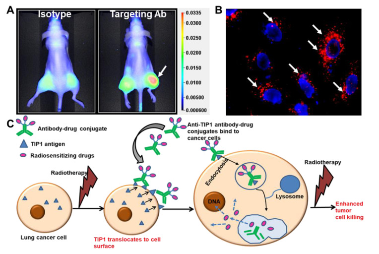
Radiation-sensitizing antibody–drug conjugates. (A) Near-infrared (NIR) imaging showing specific binding of the antibody targeting a radiation-inducible antigen to irradiated tumors (white arrow). Nude mice were injected subcutaneously with A549 cells. The right hind limb tumors were irradiated with three doses of 3 Gy. Isotype control or targeting antibody was injected via tail vein, and NIR imaging was performed to evaluate the biodistribution of the antibody (Adapted from [3]). (B) Endocytosis of ADC in cancer cells. The cancer-specific antibody was labeled with pH-sensitive dye pHrodo and incubated with cancer cells. Punctate red fluorescence indicates accumulation of the antibody in acidic compartments of the cells (white arrows). Nuclei are stained blue. (C) Schematic representation of the strategy to enhance the therapeutic efficacy of radiation and reduce detrimental side effects. Radiation enhances surface expression of radiation inducible antigens such as TIP1 in lung cancer and not normal cells, leading to specific binding of ADCs to cancer. ADCs deliver radiosensitizers to cancer. Radiotherapy then leads to enhanced cancer cell killing without effecting normal cells.

