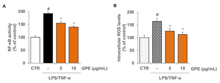
Figure 6.
Inhibitory effects of GPE on NF-κB activation and intracellular ROS levels in inflamed Caco-2 cells. Differentiated Caco-2 cells were pre-treated with various concentrations of GPE (5 and 10 µg/mL) for 2 h, followed by incubation with LPS (10 μg/mL) and TNF-α (10 ng/mL) for 2 h. NF-κB activation was assessed in nuclear proteins by an ELISA-based method measuring the DNA-binding activity of NF-κB (A). Intracellular ROS were analyzed by using carboxy-H2DCFDA staining by fluorescence plate reader (B). The white histograms correspond to the untreated control (CTR), while the colored histograms correspond to the different treated groups (black for LPS/TNF-α and orange for GPE+LPS/TNF-α). Each experiment was executed in triplicate. Data are reported as unstimulated control percentage (mean ± SD). # p < 0.01 versus control; * p < 0.05 versus LPS/TNF-α alone.

