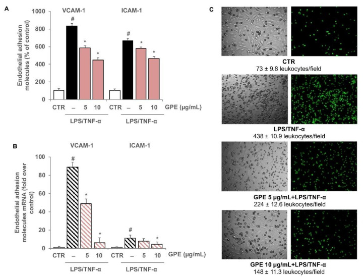
Figure 8.
Inhibitory effects of GPE on the stimulated expression of endothelial adhesion molecules and on the endothelial monocyte adhesion. Differentiated Caco-2 monolayers were grown on the upper side of the inserts and placed in proximity to HMEC-1 grown on the bottom of the wells. GPE (5 and 10 μg/mL) was added on the apical compartment for 2 h, after which LPS (10 μg/mL) and TNF-α (10 ng/mL) were applied on the apical compartment and TNF-α (10 ng/mL) on the basolateral compartment for 16 h (B) or 12 h. (A,C) Endothelial cell surface expression of VCAM-1 and ICAM-1 was assessed by cell surface EIA and expressed as percentage of unstimulated control. (B) VCAM-1 and ICAM-1 mRNA levels were analyzed by quantitative RT-PCR and expressed as fold over unstimulated control (mean ± SD). (C) HMEC-1 were co-cultured with labeled THP-1 cells for 1 h. The number of adherent THP-1 cells was monitored by fluorescence microscope or measured by the fluorescence plate reader. Each experiment was performed in triplicate. The white histograms correspond to the untreated control (CTR), while the colored histograms correspond to the different treated groups (black for LPS/TNF-α and pink for GPE+LPS/TNF-α). # p < 0.01 versus control; * p < 0.05 versus LPS/TNF-α alone.

