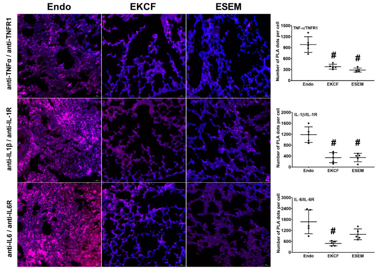
Figure 2.
Cytokine-receptor binding status. Representative microscopic images of immunofluorescence staining of proximity ligation assay (PLA) and PLA signal intensities for measuring bindings of tumor necrosis factor-α (TNFα), interleukin-1β (IL-1β), and interleukin-6 (IL-6) to the cognate receptor TNF receptor 1 (TNFR1), IL-1 receptor (IL-1R), and IL-6 receptor (IL-6R) in lung tissues in mice that were measured at 24 h after endotoxin administration. Red dots: (+) PLA signal, indicating (+) protein–protein interactions between TNF-α/TNFR1, IL-1β/IL-1R, and IL-6/IL-6R. Blue dots: 4′,6-diamidino-2-phenylindole stain, indicating a cell nucleus in lung tissues. Endo: the endotoxin group. EKCF: the endotoxin plus the KCF18 peptide group. ESEM: the endotoxin plus the SEM18 peptide group. Data were obtained from five mice in each group and presented as the mean ± standard deviation. # p < 0.05, versus the Endo group.

