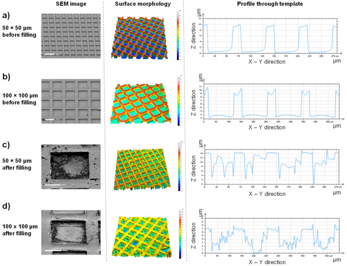
Figure 2.
Well-defined microscale templates can easily be filled with a hydrogel precursor solution. Scanning electron microscope (SEM) images (left panel) with corresponding multi-pinhole confocal microscopy height maps (middle panel) and cross-sectional data (right panel) show unfilled templates of 50 µm (a) and 100 µm (b) widths. Analysis of the filling of these templates was performed after hydrogel formation but before template removal, for both 50 µm- (c) and 100 µm- (d) width templates. Scale bars represent 100 µm (a,b), 20 µm (c), and 50 µm (d).

