Figure 1.

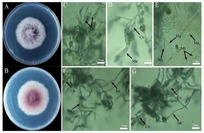
Observation of the colony morphology of strain XFT3-1 on PDA (A,B) and the conidiophore, macroconidia, and microconidia on SNA (C–G). Note: Ma: macroconidia, Mi: microconidia, Co: Conidiophore.

Observation of the colony morphology of strain XFT3-1 on PDA (A,B) and the conidiophore, macroconidia, and microconidia on SNA (C–G). Note: Ma: macroconidia, Mi: microconidia, Co: Conidiophore.