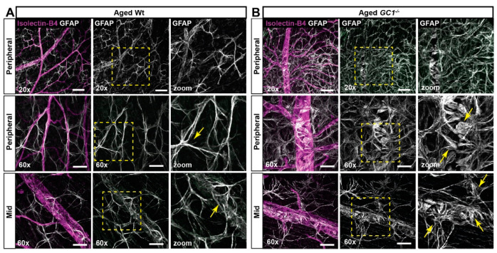
Figure 4.
Astrocyte morphology in aged Wt and GC1−/− retina. (A) Representative confocal micrographs of astrocytes (GFAP; white) and blood vessels (isolectin B4; magenta) in peripheral and mid retina of aged Wt mice. Astrocyte processes are increasingly striated and frayed (yellow arrows) in proximity to vessels compared with young mice. (B) Representative confocal micrographs of astrocytes (GFAP; white) and blood vessels (isolectin B4; magenta) in peripheral and mid retina of aged GC1−/− mice. Dense patches of matted astrocytes are observed in proximity to blood vessels at the periphery. End feet appear increasingly bulbous and frayed (yellow arrows) compared to young GC1−/− animals and Wt mice. Scale bars at 20× = 100 µm and at 60× = 40 µm.

