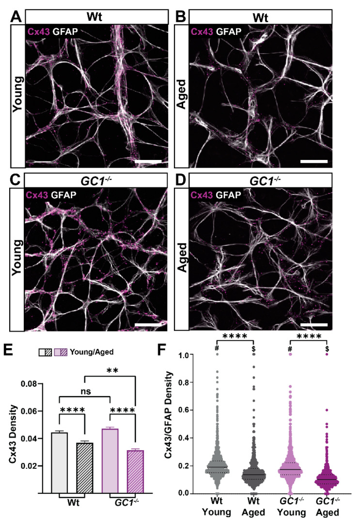
Figure 6.
Connexin 43 (Cx43) density decreases significantly with age in Wt and GC1−/− mice. (A) Representative confocal micrographs of astrocytes (GFAP; white) and Cx43 (magenta) in retina of young Wt mice. (B) Representative confocal micrographs of astrocytes (GFAP; white) and Cx43 (magenta) in retina of aged Wt mice. (C) Representative confocal micrographs of astrocytes (GFAP; white) and Cx43 (magenta) in retina of young GC1−/− mice. (D) Representative confocal micrographs of astrocytes (GFAP; white) and Cx43 (magenta) in retina of aged GC1−/− mice. Scale bar = 40 µm. (E) Cx43 density across entire retinae of young and aged Wt and GC1−/− mice. Cx43 significantly decreases with age in Wt mice (n = 8 Wt, n = 10 GC1−/−; **** p < 0.0001) and significantly decreases with age in GC1−/− mice (n = 3 Wt, n = 4 GC1−/−; **** p < 0.0001). There is a significant difference between aged Wt and GC1−/− mice (n = 8 Wt and n = 4 GC1−/−; ** p = 0.005) (F) The ratio of Cx43 to GFAP density for each quadrant analyzed shows a significant difference between young and aged Wt mice (**** p < 0.0001) and young and aged GC1−/− mice (**** p < 0.0001). There is also a significant difference between genotypes in young (# p < 0.0001) and aged ($ < 0.0001) mice. All data expressed as means ± S.E.M. Statistical analyses carried out were Kruskall–Wallis one-way ANOVA. ns: not significant.

