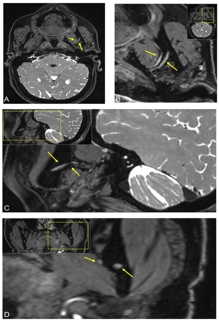
Figure 2.
Follow-up MRI after three months of the same patient. The 3D Double-Echo Steady State (DESS) MRI protocol revealed the following: (A,B) Axial, (C) sagittal, and (D) coronal side-by-side comparison of the image reconstructions showed bilateral symmetry of the mandibular nerve with a loss of signal intensity in the 3D-DESS sequence compared with the previous examination, no longer indicating evident pathological signal alterations. In all images, the short arrow shows signal hyperintensities of the lingual nerve at the anatomic point where it begins to pass from lateral to medial and the longer arrow represents the inferior alveolar nerve before it enters into the mandibular foramen. For orientation, the dotted rectangles in the corner show the enlarged area.

