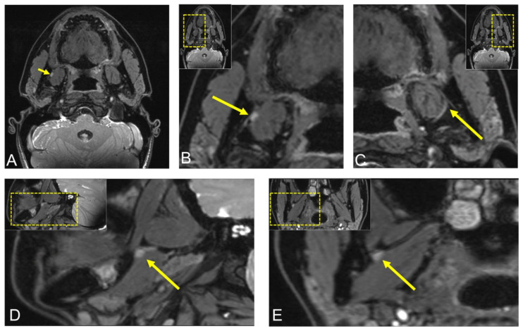
Figure 4.
Postoperative visualization of the lingual nerve (LN) using 3D double-echo steady-state (3D-DESS) MRI in the same patient. (A,B) Axial, (D) sagittal, (E) coronal reconstruction of the region of interest displaying the LN asymmetry throughout its course through the infratemporal fossa, with an LN hyperintense nodular lesion of 5 × 3 mm between the right medial pterygoid muscle and the mandible. (C) Axial image reconstruction of the healthy LN on the contralateral side for comparison. For orientation, the dotted rectangles in the corner show the enlarged area.

