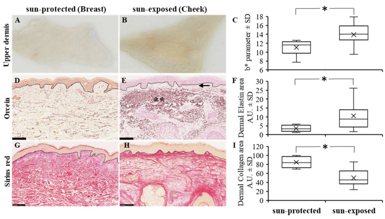
Figure 3.
Ex vivo human dermis skin (A,B), b* parameter measurement (C), and orcein (D,E) and sirius red (G,H) histological stains from sun-protected (A,D,G) and sun-exposed (B,E,H) human skin (scale bar = 100 µm). Detection of b* parameter, elastin, and collagen in dermis were increased in sun-exposed skin as compared to sun-protected skin as reported in box plot (C,F,I, respectively) (* p < 0.05); arrow and ** indicate, respectively, the grenz zone and amorphous elastic aggregate both characteristic of elastosis area in sun-exposed dermis.

