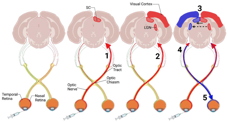
Figure 1.
Prion spread following intraocular inoculation. Inoculation of prions into the retina of rodents results in anterograde spread of prion agent from the inoculated retina to the contralateral superior colliculus (SC; 1) and lateral geniculate nucleus (LGN), and visual cortex (solid red line and red structures; 2). As the animal approaches clinical endpoint, the ipsilateral SC, LGN, and visual cortex are also affected to a lesser extent (blue structures). This is thought to be due to reciprocal connections between the contralateral SC (solid black arrow; 3) and LGN (dashed black arrow; 3) and ipsilateral anterograde spread from the inoculated retina (dashed red line; 4). The uninoculated optic nerve and retina are also affected through retrograde prion spread (solid blue line; 5). Image created with BioRender.com, accessed on 28 February 2022.

