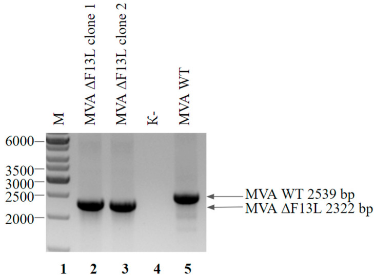
Figure 5.
PCR analysis of the MVA ΔF13L viral DNA. Genomic DNA of MVA ΔF13L from clone 1 (lane 2), clone 2 (lane 3) and wild-type MVA (lane 5) served as a template DNA. Negative control was run without any template (lane 4). Molecular weights were determined in comparison to the 1-kb ladder (lane 1).

