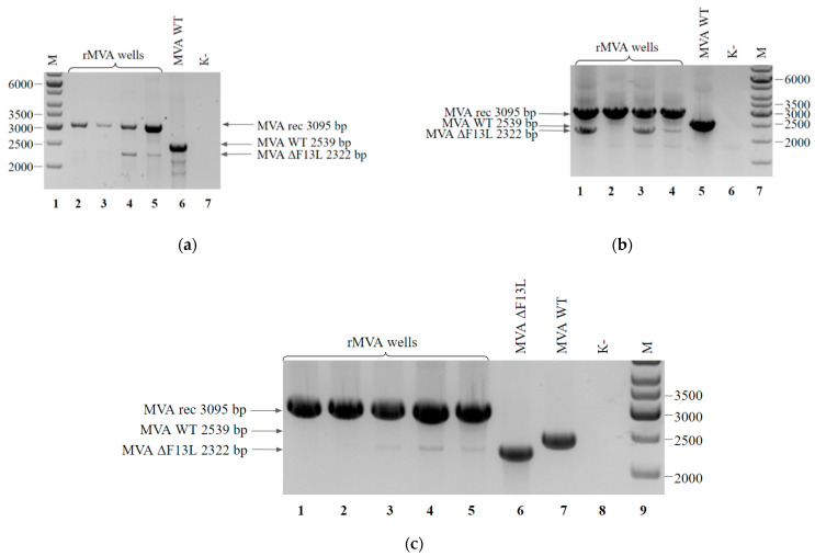
Figure 7.
PCR analysis of rMVA-F13Lrev viral DNA: (a) Clones after four blind passages and isolation by limiting dilution method. Cells from the wells contained single plaques (lanes 2–5) and wild-type MVA (lane 6) served as a template DNA. Negative control was run without any template (lane 7). Molecular weights were determined in comparison to the 1-kb ladder (lane 1); (b) Clones after one round of plaque isolation, subsequent two blind passages of selected plaques and isolation by limiting dilution method. Cells from the wells contained single plaques (lanes 1–4) and wild-type MVA (lane 5) served as a template DNA. Negative control was run without any template (lane 6). Molecular weights were determined in comparison to the 1-kb ladder (lane 7); (c) Clones after two blind passages and isolation by limiting dilution method. Cells from the wells contained single plaques (lanes 1–5), MVA ΔF13L (lane 6) and wild-type MVA (lane 7) served as a template DNA. Negative control was run without any template (lane 8). Molecular weights were determined in comparison to the 1-kb ladder (lane 9).

