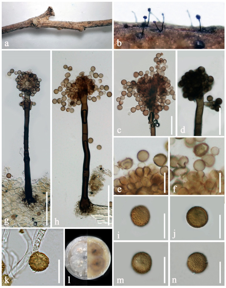
Figure 4.
Periconia byssoides (KUN-HKAS 107383). (a) Colonies on the host substrate; (b) closed up conidiophores on host; (c,d) conidial heads bearing conidiogenous cells and conidia; (e,f) conidiogenous cells with attached conidia; (g,h) conidiophores with spherical conidial heads; (i,j,m,n) conidia; (k) germinated conidia; (l) forward and reverse colonies on PDA. Scale bars: (g,h) = 100 μm, (c,d) = 50 μm, (e,f) = 20 μm, (i–k,m,n) = 15 μm.

