Figure 4.
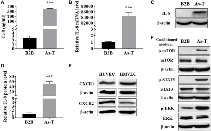
As-T cells released higher level of IL-8, which activated the IL-8 pathway in endothelial cells via the paracrine effect. (A) The protein levels of IL-8 in CM from B2B and As-T cells were measured using ELISA assay. ***p < 0.001 compared to B2B cells. (B) The mRNA level of IL-8 was measured using qRT-PCR. ***p < 0.001 compared to B2B cells. (C) The protein level of IL-8 in B2B and As-T cells was determined using immunoblotting assay. (D) Quantification of the immunoblotting results using Image Lab software. ***p < 0.001 compared to B2B. (E) The protein expression of IL-8 receptors, CXCR1 and CXCR2, in HUVECs (left) and HMVECs (right) was determined using immunoblotting assay. (F) HUVECs were starved overnight and then treated with CM from B2B or As-T cells for 6 h. The expression of IL-8/IL-8R signaling pathway molecules, p-mTOR, mTOR, p-STAT3, STAT3, p-ERK, ERK, and β-actin was analyzed using immunoblotting assay.

