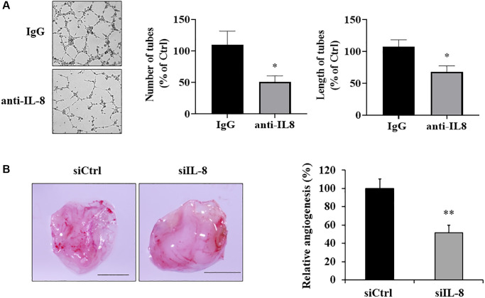
Figure 5.
IL-8 generated by As-T cells was required for angiogenesis in vitro and in vivo. (A) IgG or anti-IL-8 neutralizing antibody was added to the CM of As-T cells and incubated for 30 min, then the CM was mixed with starved HUVECs in basic EBM2 in a 1:1 ratio to perform the tube formation assay as described above. The images of the tubular structures were captured 6–12 h later using an inverted microscope at 4× magnification. Left panel: representative images of the tubular structures. Middle panel: quantification of the number of the tubular structures. Right panel: quantification of the length of the tubular structures. *p < 0.05 compared to IgG control. (B) IL-8 was knocked down in As-T cells by transfection of siRNA against IL-8, and angiogenesis assay was performed to evaluate the effects of IL-8 silencing on angiogenesis using CAM model. Left panel: representative plugs from siRNA scrambled control (siCtrl) and siIL-8 groups. Scale is 2 mm. Right panel: the number of blood vessel branches was counted from eight replicates and normalized to that of the control group. **p < 0.01 compared to the siCtrl group.

