Figure 7.
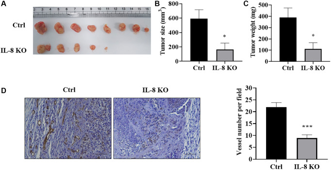
IL-8 knockout in As-T cells attenuated tumor growth and angiogenesis using HMVEC/As-T co-implantation animal model. IL-8 was knocked out in As-T cells using the CRISPR/Cas9 technique. The HMVEC cells (0.9 × 10⁶) were mixed with wild type Ctrl or IL-8 KO As-T cells (0.1 × 10⁶) in basic EBM2 medium and then mixed with 1:1 (v/v) growth factor-reduced Matrigel. The cell suspension was subcutaneously injected into flanks of nude mice at 6 weeks of age. The tumors were harvested 6 weeks after the implantation. (A) Representative tumors were shown. (B) The size of tumors. *p < 0.05 compared to the As-T Ctrl+HMVECs group. (C) The weight of the tumors. *p < 0.05 compared to the As-T Ctrl+HMVECs group. (D) IHC staining of CD31 in tumors. Left panel: representative images of IHC staining. Scale, 50 μm. Right panel: quantification of the microvessel structures in the IHC images. ***p < 0.001 compared to the Ctrl group.

