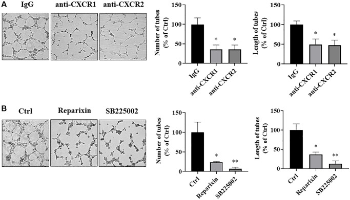
Figure 8.
IL-8 receptors in vascular endothelial cells were required for angiogenesis. (A) HUVECs were starved overnight and resuspended in basic EBM2 medium supplemented with IgG or anti-CXCR1 or anti-CXCR2 neutralizing antibodies for 30 min. Then the cells were mixed with CM from As-T cells in a 1:1 ratio and plated in 96 well plates pre-coated with solidified growth factor-reduced Matrigel for tube formation assay. The tubular structures were imaged at 6–12 h at 4× magnification. Left panel: representative images of the tubular structure. Middle panel: the number of tubular structures was measured. Right panel: the length of the tubular structures was measured. *p < 0.05 compared to the IgG control group. (B) HUVECs were starved overnight. The cells were then treated with DMSO, Reparixin (CXCR1 inhibitor), or SB225002 (CXCR2 inhibitor) for 2 h. The tube formation assay was performed as above. The tubular structures were imaged at 6–12 h using a microscope at 4× magnification. Left panel: the representative images of the tubular structure. Middle panel: quantification of the number of the tubular structures. Right panel: quantification of the length of the tubular structures. * and **p < 0.05 and p < 0.01, respectively, compared to the DMSO treatment control group.

