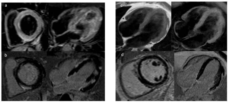
Figure 2.
In the left panel T2-STIR (a) and PSIR-TFE (b) sequences showing increased signal predominantly subepicardial patchy of the left ventricular wall due to necrosis of acute myocarditis. In the right panel sequence T1-TSE and T1-Fat Sat (c) showing multiple areas of adipose infiltration with mesocardial and subepicardial distribution of the left ventricle walls, (d) extended subepicardial signal hyperintensity in PSIR sequences for the study of “Late Gadolinium Enhancement” indicative of fibrosis for Left Dominant Arrhythmogenic Dysplasia.

