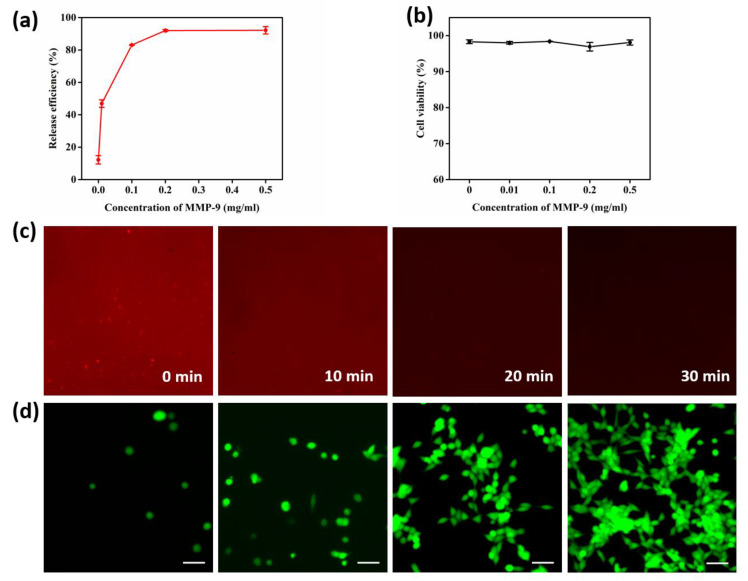
Figure 5.
Optimization of MMP-9 concentration and cell release performance. (a) Cell release efficiency treated with different concentrations of MMP-9 (0, 0.01, 0.1, 0.2 0.5 mg/mL). (b) Cell viability after treatment with different concentrations of MMP-9 for 30 min at 37 °C. (c) Fluorescence images of PE-SA-conjugated GNP substrate treated with MMP-9 for 0, 10, 20, and 30 min. (d) The viability of released cells using 0.2 mg/mL of MMP-9 solution and re-cultured at times of 0 h, 24 h, 36 h, and 48 h.

