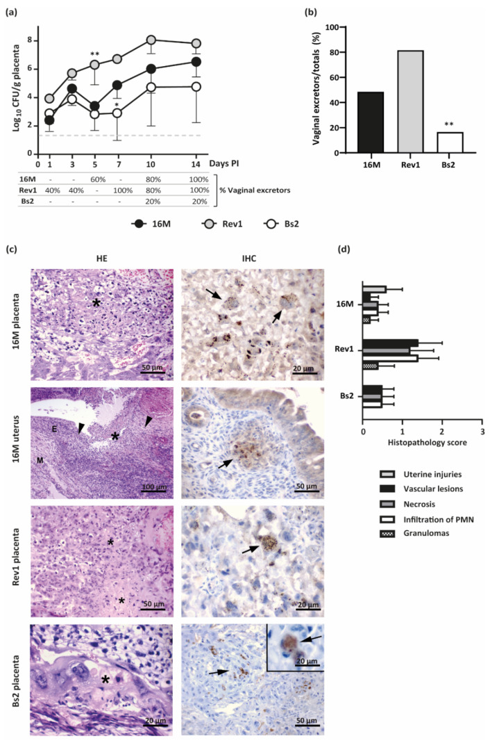
Figure 5.
Kinetics of placental infections and vaginal shedding along pregnancy and histopathological findings at 18.5 DG in mice infected by B. melitensis or B. suis bv2. (a) Placental infections (mean ± SD) and percentage of vaginal shedding along pregnancy; (b) Percentage of Brucella excretors by vaginal vs. the total pregnant mice of the study; (c) Representative images of the HE (left column) and IHC (right column) in placentas or uterus at 18.5 DG. Asterisks indicate foci of necrosis with degenerated PMN and karyorrhectic debris; arrowheads indicate uterine lesions with PMN or granulomatous infiltration; M = myometrium; E = endometrium; Arrows: Brucella antigen in TGC or in uterine epithelioid macrophages; (d) Histopathological score injuries (severity 0–3) at 18.5 DG, based on intensity and distribution of granulomas, PMN, necrosis, and vascular lesions (median ± SE). Pregnant mice were inoculated and sampled as detailed in the footnote of Figure 3. Fisher’s PLSD and Chi-square tests: * p ≤ 0.05 or ** p ≤ 0.01 vs. other groups of the point time.

