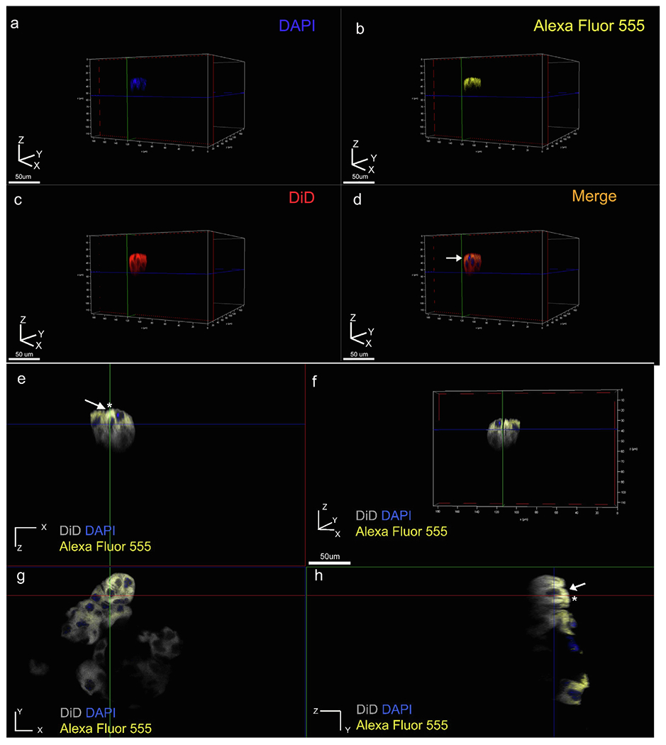
Fig. 4.

Elongated clusters of cells display polarized distribution of E-cadherin. (a-d) Three-dimensional volume rendering of a representative cell cluster viewed individual channel wise (a-c), and merged (d). Membrane labelled in red (DiD), nucleus in blue (DAPI) and E-cadherin in yellow (Alexa Fluor 555 conjugated primary antibody (yellow), (e-h) two-dimensional sectioning of representative cluster where e, g, h represents XZ, XY and ZY plane sections respectively, (f) three-dimensional volume rendering of the cluster viewed using all three channels. Membrane labeling with DiD (grey pseudo color), nucleus (blue), and E-cadherin (yellow). Membrane-cadherin co-localization is restricted to one pole of the cell along the Z axis (white arrows and asterisks in e, h).
