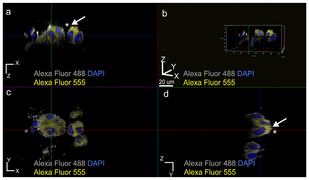
Fig. 5.

Membrane labeling using anti-actin antibody to confirm the asymmetrical E-cadherin distribution pattern within ALC clusters formed in the presence of WT recombinant AMBN. (a) XZ plane section, (b) three-dimensional volume rendering of the cluster, (c) XY plane section and (d) YZ plane section. White arrows with asterisks in a and d indicate membrane-actin colocalization pattern. Grey pseudo color for Alexa Fluor 488 actin and yellow Alexa fluor 555 conjugated anti-E-cadherin.
