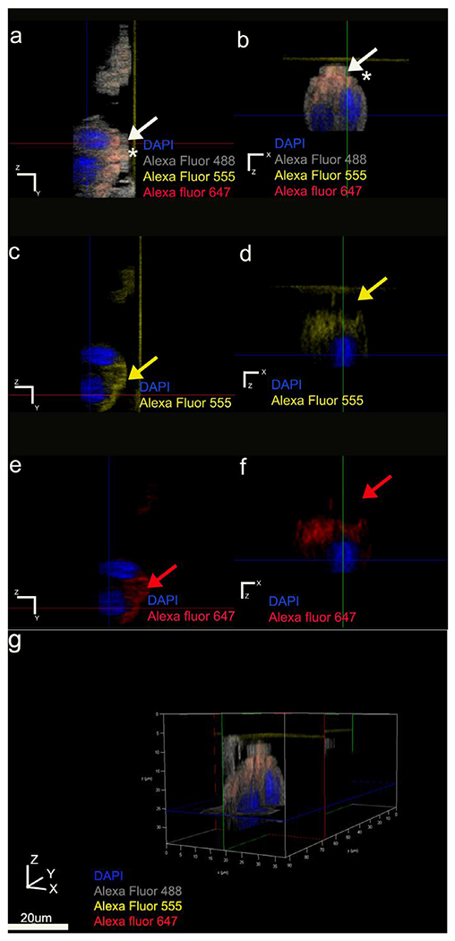
Fig. 7.

Asymmetrical immunolabeling pattern of polarity protein Par3. Par3 localization within the clusters restricted to the same pole of the cell membrane as E-cadherin. Anti-actin antibody used as the symmetrical membrane marker. Actin represented with grey pseudo color; Par3 in red, E-cadherin in yellow and nucleus in blue. (a, b) Par3 and E-cadherin colocalize (represented in orange) basal to the nucleus position indicated by white arrows and asterisks. (c, d) YZ and XZ plane sections of Alexa Fluor 555 E-cadherin channel respectively. (e, f) YZ and XZ plane sections of Alexa Fluor 647 Par-3 channel respectively. (g) three-dimensional volume rendering of the cluster viewed using all channels.
