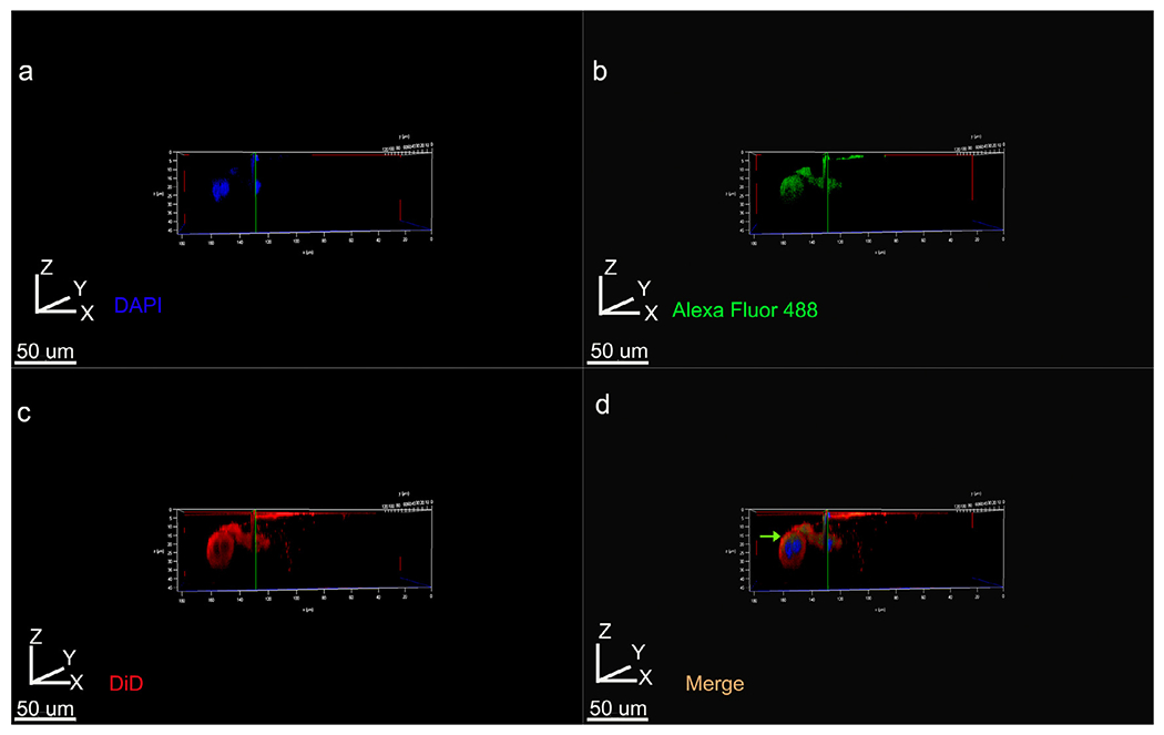
Fig. 8.

3D volume rendering of Z stacks showing tight junctional protein Claudin-1 distribution within ALC clusters labeled with DiD for cell membrane. (a) DAPI, (b) Cldn1 labeling with Alexa Fluor 488 conjugated secondary, (c) DiD, (d) merged imaged using all three channels. Cldn1 labeling is restricted to one pole of the cell and is not detectable on the below the nucleus (green arrow in d).
