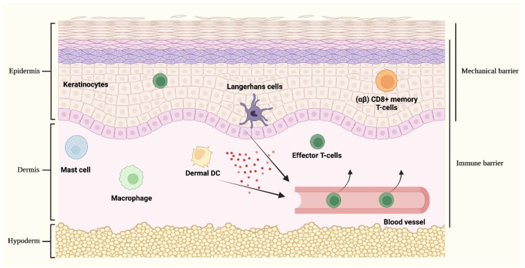
Figure 2.
Cutaneous immune responses. The skin provides a passive protective mechanical barrier tightly integrated with an active immune barrier, collectively aiming at creating a robust and intricate defence network. The mechanical barrier against external noxae is given by the highly packed keratinocytes of the epidermis. Different cellular populations act as sentinels involved in the recognition of dangerous stimuli and, upon activation, are implicated in orchestrating complex cutaneous immune responses. These cell types include keratinocytes, Langerhans cells and (αβ) CD8+ memory T-cells that are located in the epidermis, dermal dendritic cells (DCs), mast cells and macrophages. Upon stimulation, the activation of keratinocytes and epidermal/dermal resident immune cells leads to the release of cytokines and chemokines (represented by red spheres in the figure) that collectively sustain inflammatory responses and favor the recruitment of activated inflammatory cells from the bloodstream, principally of effector T-cells, hence redirecting the immune response towards the initial cutaneous inflammatory site.

