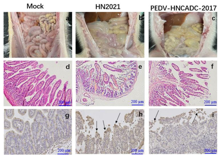
Figure 6.
Lesions and IHC of small intestinal tissue sections from piglets inoculated with two types of PEDV. (a–c) Macroscopic lesions of the small intestines from challenged and control piglets. (d–f) Microscopic damage in Jejunum in piglets (Original magnifications: ×20). (g–i) PEDV antigen detection in different intestinal tissues of piglets infected with PEDV isolates by IHC (Original magnifications: ×20). Antigens are depicted as arrows. Scale bars are shown in each picture.

