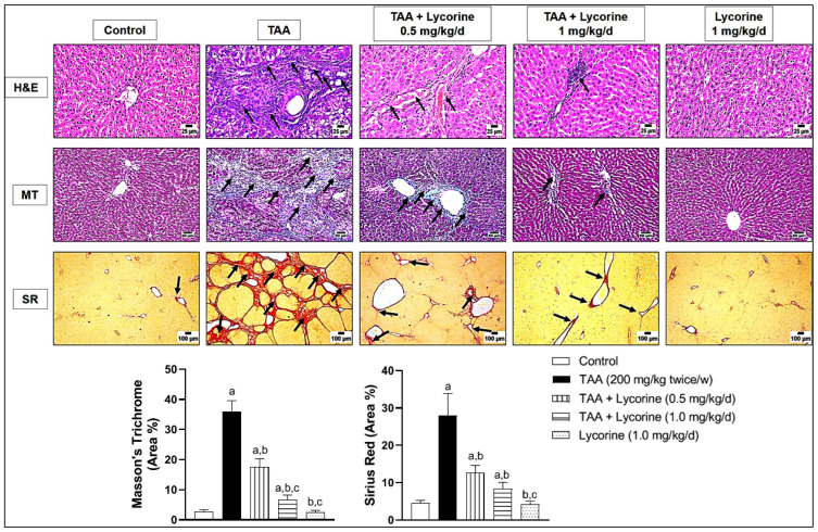
Figure 2.
Histopathological examination of liver sections stained by hematoxylin and eosin (H&E), Masson’s Trichrome (MT) and Sirius Red (SR) in a rat model of thioacetamide (TAA)-induced liver fibrosis. The control group shows a normal lobular architecture (H&E), accompanied by a normal distribution of fine collagen fibers around the central vein (MT and SR); the TAA-exposed group displays thick fibrous strands that surrounds the hepatic lobules with mononuclear inflammatory cell infiltration (arrows) and hepatocellular vacuolar degeneration (H&E), accompanied by abundant fibroplasia completely surrounding the hepatic lobules (blue with MT and red with SR); treatment with lycorine (0.5 mg/kg/d) shows a moderate improvement of the histopathological changes by all stains; treatment with lycorine (1 mg/kg/d) shows a marked improvement of histopathological changes, except for minor inflammatory cells infiltration (H&E) together with fine collagen deposition (MT & SR), as indicated by arrows; the lycorine-alone group (1 mg/kg/d) displays apparently normal hepatic tissue (H&E) and collagen deposition (MT and SR). MT and SR staining was quantified as (area %). Data are presented as mean ± S.D. (n = 6). (a), (b), (c) Statistically significant from the control, TAA, and TAA + lycorine 0.5 mg/kg/d groups, respectively, at p < 0.05 using one-way analysis of variance (ANOVA) followed by Tukey’s as a post-hoc test.

